DiagnosticApplications
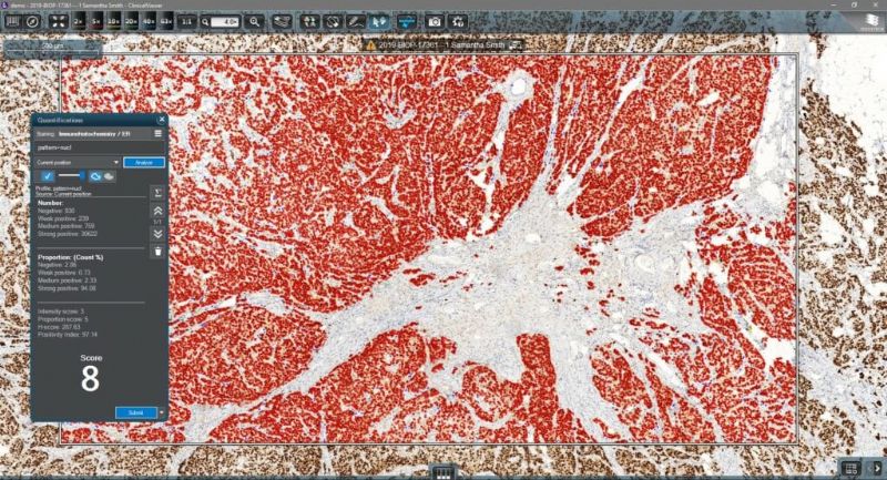
Diagnostic Applications is an image analysis platform designed as a decision support tool for fast, field of view-based, automated quantification.
With the help of this computer-aided image analysis tool, accurate, fast, and high-quality analytical results can be generated. Thanks to the integration of CaseManager and Diagnostic Applications, the measurement results, charts and masked images of the measurement area are automatically displayed in the appropriate pathology case on CaseManager’s interface. The pathologist can easily attach the measurement results and images to the final report.
The image analysis modules integrated into ClinicalViewer provide the pathologist with a quantitative image analysis framework: Four modules (Estrogen, HER2, Ki67 and Progesterone) help quantify solid tumor cases. The results of the analysis are then included in the final report in CaseManager.
Key features
- Four image analysis tools (Er, Pr, HER2, Ki67) offer image analysis solutions designed for diagnostic breast panel assessment with the IVD-approved MembraneQuant and NuclearQuant algorithms inside
- Express image analysis process on current view or predefined annotation
- Measurement result saving process: the result of the image evaluation is saved to the patient data
Further available image analysis tools
PatternQuant

PatternQuant is a trainable pattern recognition module for tissue classification, tissue pre-segmentation and identification of several tissue structures. The artificial intelligence-based algorithm is able to learn and classify various tissue types based on their texture patterns and colour features. This module can analyze fluorescent and brightfield slides.
CellQuant

The Swiss army knife of 3DHISTECH’s image analysis tools, CellQuant is a versatile cell detection application, an optimal solution for investigating various IHC stainings (best suited for Ki67 slides). The application is adequate for cell nuclei, cytoplasmic and membrane marker quantification. The software reports the results like positivity ranges of cell nuclei, cytoplasm or membrane signals. This module can analyze fluorescent and brightfield slides.
HistoQuant

HistoQuant is an image segmentation module which identifies stained tissue elements based on color and intensity features. This is an adequate solution for double-stain quantification or multiplex fluorescent analysis, with dual stain colocalization option. This module can analyze fluorescent and brightfield slides.

